Why Visual Performance Matters: Foundations for Occupational Therapy Practice — participation guide
3 contact hours · 0.3 AOTA CEUs · All modules — follow along, take notes, and complete the reflection prompts.
Visual Minds Learning · AOTA Approved Provider #0000001618
Why Visual Performance Matters: Foundations for Occupational Therapy Practice
Participation and Reflection Guide
3 contact hours · 0.3 AOTA CEUs
Print this guide before you begin. Use it to follow the content, capture what applies to your caseload, and complete the reflection prompts.
Learning outcomes
By the end of this learning experience, you will be able to:
- Explain the developmental and anatomical basis of the visual system and articulate the occupational therapy practitioner's role in supporting visual function within the OT scope of practice.
- Differentiate ventral ("what") and dorsal ("where") visual pathway functions and identify classroom-based behavioral signatures associated with dysfunction in each pathway.
- Recognize clinical signs of visual conditions warranting referral to optometry, ophthalmology, or developmental vision specialists and distinguish them from findings addressable through OT intervention.
- Analyze comorbidity patterns between visual function deficits and autism, ADHD, dyslexia, and dysgraphia to inform integrated, occupation-based intervention planning.
- Apply a low-vision service delivery model, including environmental modification strategies, to support occupational performance for clients with reduced visual function.
- Construct an immediate next-step plan for a client with suspected visual performance limitations, sequencing clinical observation, occupation-based documentation, referral, and structured follow-up assessment within the OT scope of practice.
Reflect: how will achieving these outcomes help you meet the needs of your students or clients?
Agenda
- Block 1 — Vision as a performance system: why a passed screening tells you little · 25 min
- Block 2 — What vision looks like in occupation: classroom, clinic, and home signatures · 30 min
- Block 3 — Recognize and refer: red flags by urgency and the scope line · 35 min
- Block 4 — When it is not just vision: autism, ADHD, dyslexia, dysgraphia · 30 min
- Block 5 — Modify Monday morning: environmental and task modifications · 35 min
- Block 6 — Where this goes next: from observation to structure and measurement · 25 min
Module 1
Why Visual Performance Matters
Anchor concept
Vision is a performance system, not a single measurement. A passed acuity screening tells you the student can resolve a letter at distance — nothing about whether they can use vision to do the work of the day.
Follow along
What this module covers
- You are already treating vision
- Understand → assess → intervene
- Vision is the dominant channel for learning
- Why schools are required to respond
- Where OT sits relative to the eye care team
Key ideas
Capture these as you listen
- Visual performance is already on your caseload; the only question is whether it is named and measured.
- Understanding without assessment is a guess; assessment without intervention is abandonment.
- Vision is a gateway system; visual deficits appear as reading, writing, mobility, and participation problems.
- Typical visual development follows a predictable timeline; persistent gaps past those windows warrant referral.
- OTs describe functional visual performance and occupational impact; eye care providers make the medical diagnosis.
- IDEA and Section 504 both welcome OTs' functional, performance-based evidence.
Notes
My notes for this module
Reference figures
Keep these for practice
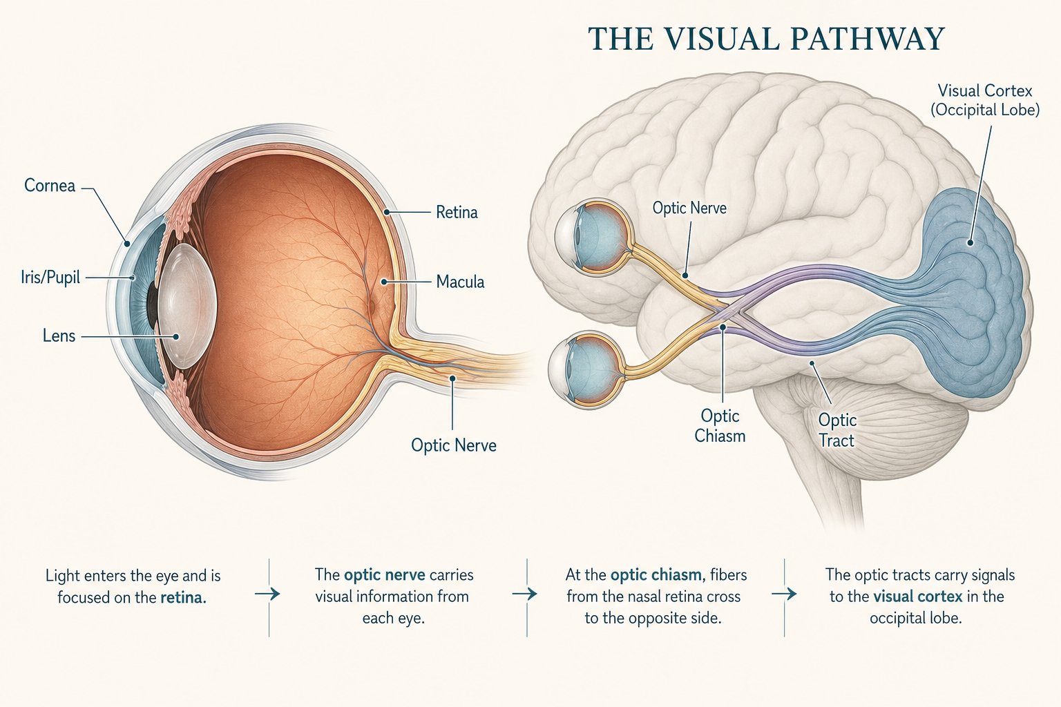
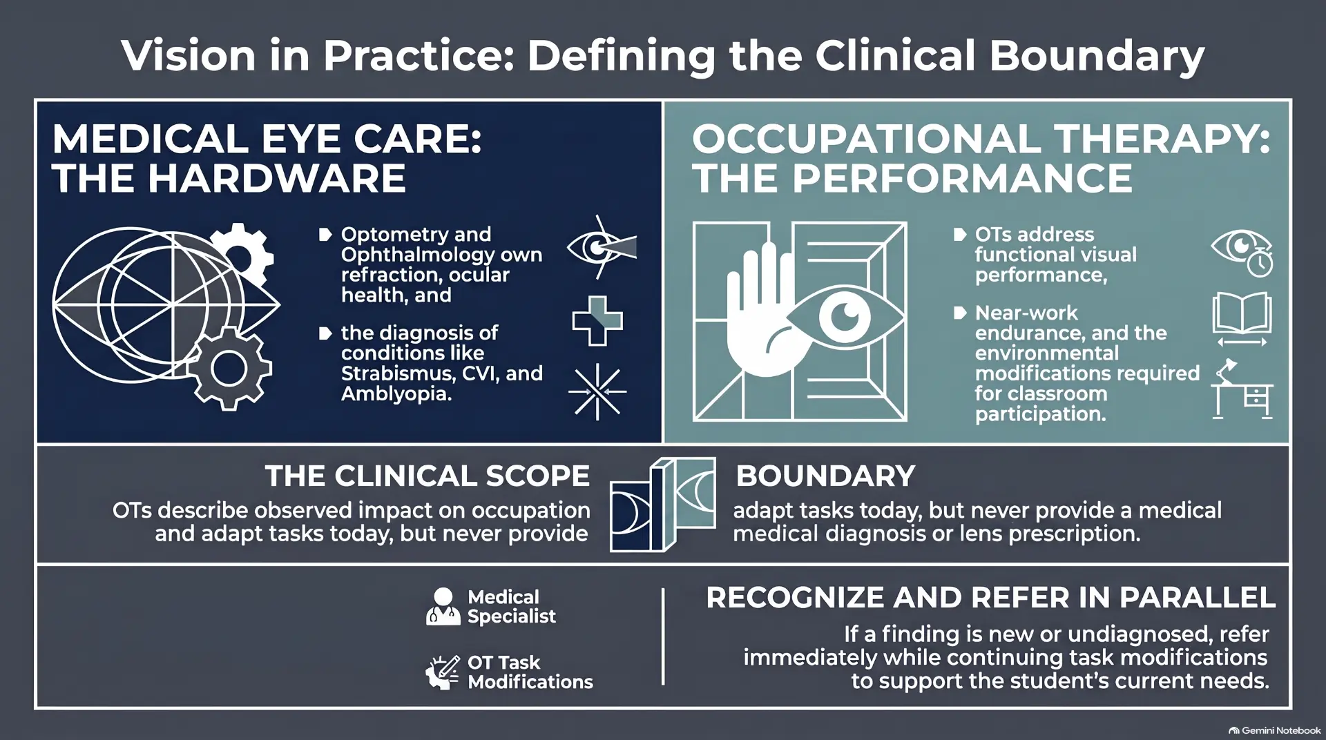
Where the referral sits in the student’s day
Apply it
Case prompt
A kindergarten teacher reports a student "just isn't paying attention" during calendar time and bumps into classmates during transitions. The school vision screen was passed six months ago. What visual-performance questions would you ask before assuming this is attention or behavior?
Practice
Mini exercise
In one sentence, describe the OT role in vision without using the words "diagnose," "therapy," or "eye doctor."
Take it to work
Action step this week
This week, write one observation of a student using only the language of functional visual performance and occupational impact. Avoid any diagnostic or medical terminology.
Module 2
The Visual Pathways
Anchor concept
Different visual jobs travel different routes. Identifying what something is and knowing where it is in space are separate demands, and students can be strong in one and weak in the other.
Follow along
What this module covers
- What a ventral-stream (parvocellular) signature looks like in class
- What a dorsal-stream (magnocellular) signature looks like in class
- We do not treat tracts — we treat the functional components
Key ideas
Capture these as you listen
- Ventral stream = "what" (object identity, detail, recognition); dorsal stream = "where" (spatial mapping, posture, movement guidance).
- Classroom signatures differ: ventral problems look like recognition errors; dorsal problems look like spatial, navigation, and postural errors.
- OTs describe the behavioral signature in occupational terms, not as a neurological diagnosis.
- Both streams feed Level 3 of a structured visual performance framework; observations must be tied to functional performance.
Notes
My notes for this module
Reference figures
Keep these for practice
Two streams, one retina
Parvocellular and magnocellular tracts
| Parvocellular | Magnocellular | |
|---|---|---|
| Cell body size | Small (parvo = “small”) | Large (magno = “large”) |
| LGN layers | Layers 3–6 | Layers 1–2 |
| Best at | Fine spatial detail, color (red/green) | Motion, low contrast, coarse form |
| Temporal response | Sustained, slower conduction | Transient, fast conduction |
| Contrast sensitivity | Needs higher contrast | Responds to very low contrast |
| Feeds primarily | Ventral “what” stream | Dorsal “where/how” stream |
| Functional signature | Slow letter/word ID, detail errors, color-cue reliance | Place loss, spatial/postural errors, motion-guided reach errors |
Vision's Two Highways
Apply it
Case prompt
A third-grade student reads words accurately in isolation but cannot find her place after looking up from the page and frequently loses items on a busy desk. Which stream pattern does this sound more consistent with, and how would you phrase it for the teacher without using stream terminology?
Practice
Mini exercise
Write a one-sentence present-level statement that names a stream-related observation and ties it to a Level 3–5 classroom task.
Take it to work
Action step this week
Pick one student on your caseload. List three visual observations and sort them by whether they look like "what" (identification) or "where" (spatial/movement) problems.
Module 3
Underlying Conditions vs. Functional Limitations
Anchor concept
The diagnosis belongs to medicine. The functional limitation belongs to you. This module is about knowing which eye and brain conditions may be sitting underneath what you are watching, so you can name the functional problem accurately instead of guessing at a cause.
Follow along
What this module covers
- How to use this module
- Naming the direction — vocabulary, not diagnosis
Key ideas
Capture these as you listen
- The diagnosis names the mechanism; the functional limitation names the OT target. Document the second, not the first.
- CVI is brain-based and often pairs with a normal eye exam; ONH, albinism, nystagmus, strabismus, amblyopia, ROP, RP, cataract, and refractive/vergence disorders change the sensory floor.
- Each condition maps to functional problems OT treats: print size, contrast, working distance, lighting, scanning, endurance, and array complexity.
- OT observation cannot rule an eye condition in or out — "see this and refer" is the wrong model; describe function and recommend an exam when none is documented.
- A short red-flag list (sudden vision loss, pain, new diplopia, unequal pupils, new turn after age 5, trauma, halos/flashes) still warrants prompt medical eye care.
Notes
My notes for this module
Apply it
Case prompt
A second-grade student with a known history of prematurity looks away from the smart board, prefers bright yellow objects, and needs repeated prompts to look at a visual target. Which conditions could plausibly sit underneath this picture, and how would you write the functional limitation you are going to treat?
Practice
Mini exercise
Name three conditions in this module that are commonly missed on a school vision screen, and explain why each one is missed.
Take it to work
Action step this week
Take two students on your caseload with a documented eye diagnosis. For each, write the diagnosis on the left and the two functional limitations you are actually treating on the right.
Module 4
Comorbidity Patterns
Anchor concept
Visual performance findings rarely arrive alone. The same child often carries an attention, learning, or developmental diagnosis, and the visual piece gets absorbed into that label.
Follow along
What this module covers
- Association is not causation
- Visual performance signs associated with dysgraphia
- The numbers behind the urgency
Key ideas
Capture these as you listen
- Autism commonly co-occurs with visual attention and ocular motility concerns; do not assume visual findings are "just autism."
- ADHD can coexist with genuine visual inefficiency; test whether reducing visual demand improves performance.
- Dyslexia is language-based, not vision-based; OTs avoid unproven visual cures and support occupational impact instead.
- Dysgraphia is strongly linked to visual-motor and spatial-planning deficits — a core OT intervention area.
- Comorbid plans are integrated, but scope boundaries remain clear: OT treats performance, eye care treats ocular health.
Notes
My notes for this module
Apply it
Case prompt
A student with ADHD and dyslexia complains that reading makes his eyes "tired." The school team wants to try a colored overlay first. How do you respond in a way that respects evidence and keeps the student's occupational needs in focus?
Practice
Mini exercise
Write a one-paragraph team note that distinguishes dyslexia from a possible co-occurring visual efficiency problem, using correct scope language.
Take it to work
Action step this week
Review one IEP for a student with a diagnosis from this module. Identify one visual performance question that has not been addressed, and one sentence you would add to the present levels.
Module 5
The Low-Vision Service Model
Anchor concept
Adaptation is intervention. You do not wait for the eye exam to make the task usable.
Follow along
What this module covers
- Two categories of clients drive two different plans
- Lighting is a modifiable variable, not a fixed constraint
- Low vision: what OT addresses, and when OT refers
- Organization, mobility, and multisensory support
Key ideas
Capture these as you listen
- The low-vision model has four levels: increase visibility, enhance with magnification, augment with another channel, substitute with another channel.
- Environmental modifications (lighting, glare, contrast, size, typeface, seating) are core OT interventions.
- Modifications must be selected for a specific occupation and goal, not applied generically.
- OTs do not diagnose low vision or prescribe optical devices; we partner with eye care providers and document functional outcomes.
Notes
My notes for this module
Reference figures
Keep these for practice
What those contrast values actually look like
Circle the word that names the picture.
~90% contrast — Black on white — target for worksheets and labels.
Circle the word that names the picture.
~70% contrast — Typical newspaper / textbook print.
Circle the word that names the picture.
~40% contrast — Faded photocopy — many classroom handouts land here.
Circle the word that names the picture.
~10% contrast — Low-contrast worksheet; effectively inaccessible.
Circle the word that names the picture.
Reverse contrast — white on dark; often preferred with glare or photophobia.
Circle the word that names the picture.
Tinted background — trial it; keep it only if performance measurably improves.
Magnification options, side by side
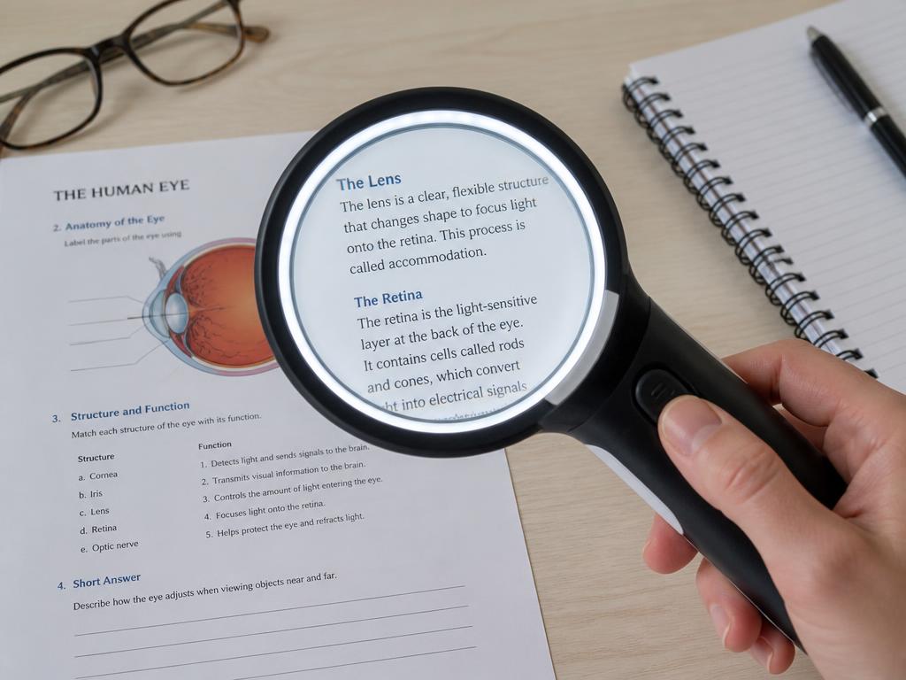
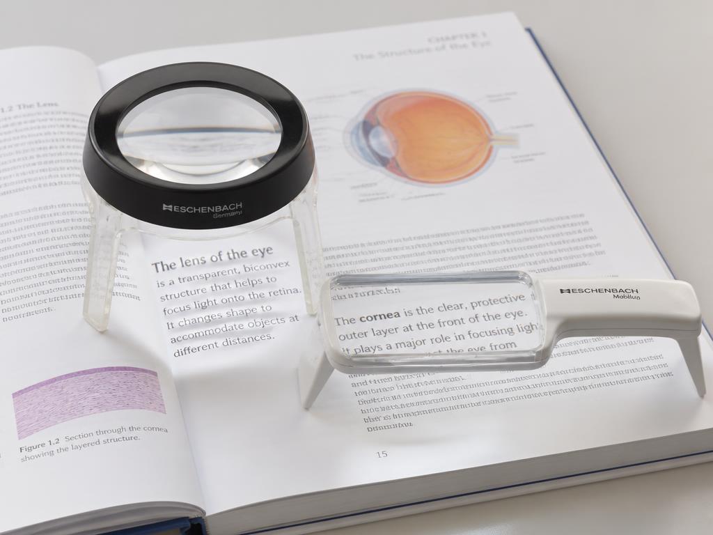

Handheld magnifier
Short spot tasks: price tags, labels, a single line of text.
Portable and cheap; requires a steady hand and short focal distance.
Stand magnifier
Sustained near work for students with tremor or poor motor control.
Fixed focal distance holds the lens steady; less portable, needs desk space.
Hands-free / spectacle
Writing and bimanual tasks where both hands must stay free.
Prescribed by the eye care team; very short working distance at high power.
Telescope (mono/binocular)
Distance targets: the board, signage, a demonstration across the room.
Spot use only — narrow field, not for travel or continuous viewing.
Electronic / video magnifier (CCTV)
Long reading and writing sessions; textbooks, worksheets, science labs.
Adds adjustable magnification, reverse contrast, and speech; cost and setup.
Built-in digital magnification
Phone/tablet zoom, text enlargement, VoiceOver, screen readers.
Already in the student’s hands and free; teach it before purchasing hardware.
Where low vision sits in the framework
Module 1 — Vision as a gateway system
Frames the whole system; every level is introduced.
Module 2 — Visual pathways (ventral / dorsal)
Magnocellular/dorsal work shows at Level 2 efficiency; parvocellular/ventral identification is Level 3 processing.
Module 3 — Underlying Conditions vs. Functional Limitations
Ocular and neurological conditions act on the sensory foundation and on binocular efficiency; OT treats the functional limitation, not the diagnosis.
Module 4 — Comorbidity patterns
Autism, ADHD, dyslexia, dysgraphia signatures appear as processing and visual-motor integration performance.
Module 5 — Low-vision service model
Low vision is a Level 1 sensory-foundation problem. Contrast, lighting, and magnification restore access at the floor; the Level 5 change is the downstream result, not the target.
Module 6 — Where this goes next
Functional integration is where all findings resolve into a plan of care.
Apply it
Case prompt
A high school student with albinism needs to read a textbook and navigate the hallway. Name one modification for each of the four low-vision levels for each goal.
Practice
Mini exercise
Choose one low-vision level and write a one-sentence accommodation for a student in your caseload or a hypothetical case.
Take it to work
Action step this week
Walk through one classroom in your building and list three environmental changes that would increase visibility for a student with reduced acuity or contrast sensitivity.
Module 6
Where This Goes Next
Anchor concept
You now have observation, referral, and modification. What you do not yet have is a repeatable structure for deciding what to look at, in what order, and how to defend the plan.
Follow along
What this module covers
- Five blocks in, here is the skill set
- Observation alone is unordered
- What the VPA™ adds on top of structure
Key ideas
Capture these as you listen
- Observation is the entry skill; without structure it is unordered and hard to defend to a team.
- A level-based organization of visual performance tells you where to look first, what a finding means, and how to sequence targets in documentation.
- Standardized administration and scoring turn a clinical impression into a comparable, re-measurable baseline.
- Every participant leaves with one described student, one referral decision, and one modification with an observable check.
Notes
My notes for this module
Reference figures
Keep these for practice
Which level each block of this course was operating on
Module 1 — Vision as a gateway system
Frames the whole system; every level is introduced.
Module 2 — Visual pathways (ventral / dorsal)
Magnocellular/dorsal work shows at Level 2 efficiency; parvocellular/ventral identification is Level 3 processing.
Module 3 — Underlying Conditions vs. Functional Limitations
Ocular and neurological conditions act on the sensory foundation and on binocular efficiency; OT treats the functional limitation, not the diagnosis.
Module 4 — Comorbidity patterns
Autism, ADHD, dyslexia, dysgraphia signatures appear as processing and visual-motor integration performance.
Module 5 — Low-vision service model
Low vision is a Level 1 sensory-foundation problem. Contrast, lighting, and magnification restore access at the floor; the Level 5 change is the downstream result, not the target.
Module 6 — Where this goes next
Functional integration is where all findings resolve into a plan of care.
Apply it
Case prompt
Take the student you have had in mind through this course. Write the referral-or-not decision in one sentence, the modification you will make in one sentence, and the observable indicator you will watch in one sentence. Would a colleague reading only those three sentences know what you saw and why you acted?
Practice
Mini exercise
In one sentence, state what a structured, level-based organization of visual performance would give you that your current observation notes do not.
Take it to work
Action step this week
Before your next treatment day, put one modification from Module 5 in place for one student and record the single observation you will use to judge whether it helped.
Video and demonstration reflections
Use this page for any video, demonstration, or case walkthrough in the course.
What did you notice about the clinician and the student?
How could you use this with a student on your caseload?
Would you do anything differently?
What questions do you still have?
Wrap-up reflections
Use this space to note:
- Strategies to try with your students next week
- Resources or materials to explore
- Information to share with your team
- Anything you want to remember